The study has shown the feasibility of this technique for food science applications, where the food’s microstructure is crucial in determining quality.
As the food sample is unaltered prior to examination, the method may well prove useful in the product design phase, where understanding product qualities or flaws is essential.
The technique, known as ptychographic X-ray computed tomography (PXCT), creates a 3D structure of a palm kernel-based oil-in-water emulsion. This had so far not been possible using other techniques.
PXCT is a 3D nano-imaging technique that offers a spatial resolution in the 100 nanometre range. It provides quantitative information by reconstructing the full 3D electron density distribution of the specimen.
The technique is well-suited for in-situ measurements as it allows for sample environments at room temperature and ambient pressure.
Although the availability of PXCT is limited to large-scale synchrotron facilities, commercial X-ray phase-contrast micro-CT scanners are available which could be used in the R&D departments of the food industry.
PXCT in food R&D

For the study, researchers from the University of Copenhagen and the Paul Scherrer Institute in Switzerland used a cream based on vegetable fat.
The sample used exhibited properties similar to the structures of food like cheese, yoghurt, ice cream, spreads, but also solid chocolate. The sample with the cream cheese-like system that the scientists X-rayed was about 20 microns thick.
All these products contain liquid water or fat as well as small particles of solid materials, which stick together and form three-dimensional structures.
In cheese and yoghurt the casein particles form the network. In chocolate it is the fat crystals and in ice cream and whipped cream it is the fat globules. The make-up of these foods contributes to their consistency and pleasant taste.
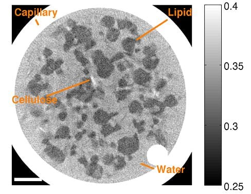
The scientists found that 98% of the fat globules in the cream cheese-like food system were cemented together in a continuous 3D network.
The different densities of food components, such as water and fat, means they have different electron densities (the number of electrons per volume).
Areas with higher electron density appeared lighter, meaning water appeared light grey. Fat appeared dark grey and the glass around the sample with a high density are seen as a white ring.
The team believe that an electron density scale could then be used to identify the various food components and study their location and structure.
"There is still a lot we don’t know about the structure of food, but this is a good step on the way to understanding and finding solutions to a number of problems dealing with food consistency, and which cost the food industry a lot of money," says asssociate professor Jens Risbo from the Department of Food Science at the University of Copenhagen.
“It's about understanding the food structure and texture. If you understand the structure, you can change it and obtain exactly the texture you want," he added.
This study represents a first for the use of PXCT in 3D imaging food products. Previously, it had only been applied for an in-situ study of water uptake in a single silk fibre.
In recent years, X-ray phase-contrast computed tomography (CT) has emerged as the imaging method of choice having been successfully applied to study the microstructure of a range of food products.
A complex food system

Commenting on their findings, the scientists say that the vegetable-based cream consisted of several ingredients. As well as water and vegetable fat, the cream also contained milk protein, stabilisers and emulsifiers.
By adjusting the addition of emulsifiers, the team believe it was possible to achieve a state in which the cream continues to be fluid until you whip it to foam, whereby all the fat globules are then reorganised and sticking together on the outside of the air bubbles in a three-dimensional system.
"It is a difficult balance, because you only want the fat globules to stick together when the cream is whipped - not if it is simply being exposed to vibration or high temperatures,” said postdoctoral researcher Merete Bøgelund Munk, Department of Food Science, University of Copenhagen.
“When the fat globules nevertheless begin to stick together prematurely - for example due to too many shocks during transport - the cream will get a consistency reminiscent of cream cheese. It becomes a relatively hard lump that can be cut."
The team believed this network structure captures a lot of water, a characteristic that is shared with foods with similar network systems, which contain a solid element immersed in a liquid, invariably water.
“This applies to all semi-solid and solid products such as chocolate, butter, cheese and spreads. The network of the cream cheese-like system is thus a model for something general in our food," commented Risbo.
The structure and the networks of these foods have remained a mystery up until the use of PXCT in the study. Previous investigations could only ever view the surface or only slightly underneath the surface of the food material on a micron scale. In addition, images were only two-dimensional.
"If we eventually come to understand the structure of chocolate, we can change it and obtain exactly the consistency that we want. A lot of money is wasted because the consistency of chocolate is really hard to control, so the end product is not good enough and must be discarded,” said Risbo.
“A possible future understanding of the crystal network in chocolate might mean that we will be able to develop components that prevent the chocolate from becoming grey and crumbly, and thus unsaleable. It is certainly a possibility that tomographic methods could be developed so we would be able to understand the mysteries of chocolate."
Source: Food Structure
Published online ahead of print, doi:10.1016/j.foostr.2016.01.001
“Ptychographic X-ray computed tomography of extended colloidal networks in food emulsions.”
Authors: Mikkel Schou Nielsen, Merete Bøgelund Munk, Ana Diaz, Emil Bøje Lind Pedersen, Mirko Holler, Stefan Bruns, Jens Risbo, Kell Mortensen, Robert Feidenhans’l
